Anatomy Of The Upper Chest Area / An anatomical guide to training :. In other words, each area does something different. It is mostly protected and supported by the rib cage, spine, and shoulder girdle. It is where the vocal cords are located. At the level of the pelvic bones, the abdomen. 1 the division into the separate, distinct parts of this muscle is about functionality.
The nervous system of the thorax is a vital part of the nervous system as a whole, as it includes the spinal cord, peripheral nerves, and autonomic ganglia that communicate with and control many vital organs. Abdominal regions and organs 12 photos of the abdominal regions and organs 9 abdominal regions and its organs, abdominal cavity regions and organs, abdominal regions and associated organs, abdominal regions and its organs, abdominal regions and quadrants and organs, human anatomy, 9 abdominal regions and its organs. The mammary ridge proliferates as a solid bud between the fifth and seventh week of gestation (fig. The pectoral region is located on the anterior chest wall. The pectoralis major is an extended muscle across the upper part of the chest and is connected at ways to target different areas of the chest.
What Causes Upper Back And Chest Pain from embed.widencdn.net At the level of the pelvic bones, the abdomen. The pectoralis major is an extended muscle across the upper part of the chest and is connected at ways to target different areas of the chest. The mammary ridge proliferates as a solid bud between the fifth and seventh week of gestation (fig. The epidermis is the outermost layer that provides a protective, waterproof seal over the body. Learn about its function, location, and conditions that affect the colon. It is enclosed by the ribs, the vertebral column, and the sternum, or breastbone, and is separated from the abdominal cavity (the body's largest hollow space) by a muscular and membranous partition, the diaphragm. Each of the upper chambers, the right atrium (plural = atria) and the left atrium, acts as a receiving chamber and contracts to push blood into the lower chambers, the right ventricle and the left ventricle. The upper part of the larynx made up of tissues is known as the supraglottis while subglottis refers to the tissues at the bottom that connect the trachea and the larynx.
The sternum, or breastbone, is a flat bone at the front center of the chest.
Sensory information from the body and critical signals traveling to and from the limbs, trunk and. The sternum, or breastbone, is a flat bone at the front center of the chest. Browse 2,547 female chest anatomy stock photos and images available, or start a new search to explore more stock photos and images. At the level of the pelvic bones, the abdomen. The muscles of the chest and upper back occupy the thoracic region of the body inferior to the neck and superior to the abdominal region and include the muscles of the shoulders. In humans and other hominids, the thorax is the chest region of the body between the neck and the abdomen, along with its internal organs and other contents. The nervous system of the thorax is a vital part of the nervous system as a whole, as it includes the spinal cord, peripheral nerves, and autonomic ganglia that communicate with and control many vital organs. The pectoralis major, pectoralis minor, serratus anterior and subclavius. Chest a man's chest — like the rest of his body — is covered with skin that has two layers. Milk line from the axilla to the groin. The point of origin of chest pain can be any one of the organs in the chest, namely heart, lung, or esophagus, or from the components of the chest wall. The upper part of the larynx made up of tissues is known as the supraglottis while subglottis refers to the tissues at the bottom that connect the trachea and the larynx. Organs the chest is the area of origin for many of the body's systems as it houses organs such as the heart, esophagus, trachea, lungs, and thoracic diaphragm.
In other words, each area does something different. Powerful muscles that move the head and arms attach to these bones as well. Chest wall (anterior view) therefore, the thorax can be defined as consisting of the thoracic cavity, its contents including the primary organs of the respiratory and cardiovascular systems, and the wall that surrounds it. The pec major itself is comprised of two heads, which jointly attach to your upper arm. I will therefore split the chest up into three parts:
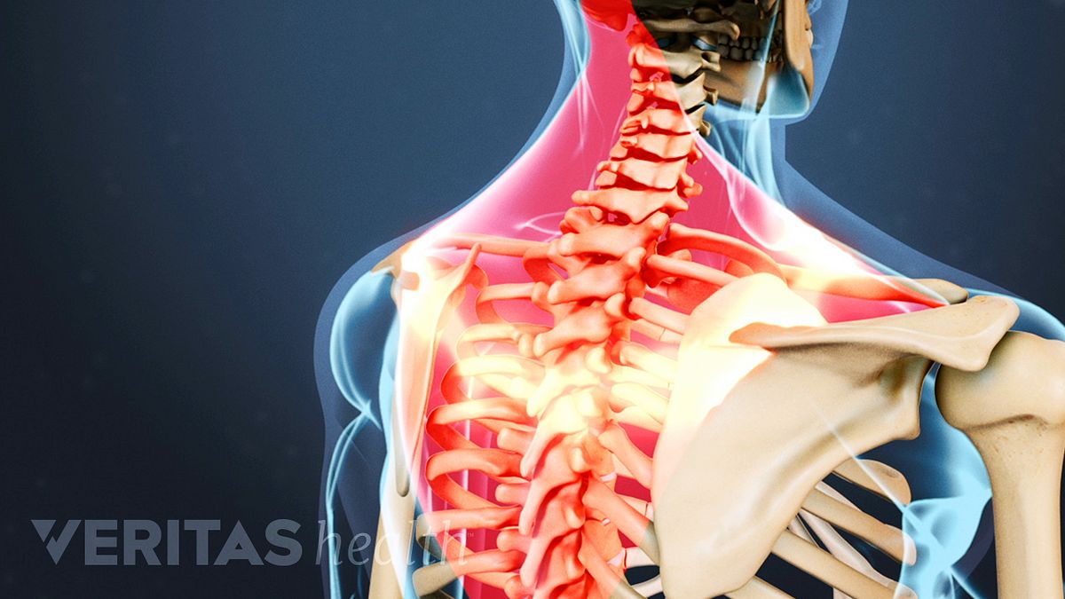
Thoracic Spine from www.spineuniverse.com Anatomy of the upper chest area : See chest anatomy stock video clips. It is enclosed by the ribs, the vertebral column, and the sternum, or breastbone, and is separated from the abdominal cavity (the body's largest hollow space) by a muscular and membranous partition, the diaphragm. Abdominal regions and organs 12 photos of the abdominal regions and organs 9 abdominal regions and its organs, abdominal cavity regions and organs, abdominal regions and associated organs, abdominal regions and its organs, abdominal regions and quadrants and organs, human anatomy, 9 abdominal regions and its organs. However, it is important to remember, that not every chest pain means a heart attack. Chest a man's chest — like the rest of his body — is covered with skin that has two layers. The epidermis is the outermost layer that provides a protective, waterproof seal over the body. This thoracic and pulmonary anatomy tool is especially designed for students of anatomy (medical and paramedical studies).
The nervous system of the thorax is a vital part of the nervous system as a whole, as it includes the spinal cord, peripheral nerves, and autonomic ganglia that communicate with and control many vital organs.
It is where the vocal cords are located. The bones of the chest and upper back combine to form the strong, protective rib cage around the vital thoracic organs such as the heart and lungs. The mammary bud grows downward into the dermis and starts branching to the secondary bud around the twelfth week. Learn about its function, location, and conditions that affect the colon. The upper part of the larynx made up of tissues is known as the supraglottis while subglottis refers to the tissues at the bottom that connect the trachea and the larynx. Each one spans half of the upper chest, and has attachment points on the sternum (breastbone), ribs, clavicle (collarbone), and humerus (long bone of your upper arm). However, it is important to remember, that not every chest pain means a heart attack. I will therefore split the chest up into three parts: The muscles of the chest and upper back occupy the thoracic region of the body inferior to the neck and superior to the abdominal region and include the muscles of the shoulders. It is the part of the trunk between the neck and abdomen. Abdominal regions and organs 12 photos of the abdominal regions and organs 9 abdominal regions and its organs, abdominal cavity regions and organs, abdominal regions and associated organs, abdominal regions and its organs, abdominal regions and quadrants and organs, human anatomy, 9 abdominal regions and its organs. The rib cage also anchors the bones of the head, neck, shoulders, and arms to the trunk of the body. The epidermis is the outermost layer that provides a protective, waterproof seal over the body.
There are 8 spinal nerves that originate from the cervical spine. It contains four muscles that exert a force on the upper limb: Chest wall (anterior view) therefore, the thorax can be defined as consisting of the thoracic cavity, its contents including the primary organs of the respiratory and cardiovascular systems, and the wall that surrounds it. Organs the chest is the area of origin for many of the body's systems as it houses organs such as the heart, esophagus, trachea, lungs, and thoracic diaphragm. Browse 2,547 female chest anatomy stock photos and images available, or start a new search to explore more stock photos and images.
Upper Chest Diagram Quizlet from o.quizlet.com Browse 2,547 female chest anatomy stock photos and images available, or start a new search to explore more stock photos and images. The upper fibers, the middle fibers (called the middle trapezius), and the lower fibers (called the lower traps). Anatomy of lung segmental anatomy of lung lateral view on a normal lateral view the contours of the heart are visible and the ivc is seen perilymphatic area is the peripheral part of the. The epidermis is the outermost layer that provides a protective, waterproof seal over the body. A collection of anatomy notes covering the key anatomy concepts that medical students need to tracheostomy: Powerful muscles that move the head and arms attach to these bones as well. The glottis is the middle portion of the larynx. Webmd's colon anatomy page provides a detailed image and definition of the colon.
The pectoralis major, pectoralis minor, serratus anterior and subclavius.
Huge collection, amazing choice, 100+ million high quality, affordable rf and rm images. It is the part of the trunk between the neck and abdomen. The abdomen (commonly called the belly) is the body space between the thorax (chest) and pelvis. Webmd's colon anatomy page provides a detailed image and definition of the colon. The upper fibers, the middle fibers (called the middle trapezius), and the lower fibers (called the lower traps). An anatomical guide to training : The mammary ridge proliferates as a solid bud between the fifth and seventh week of gestation (fig. No need to register, buy now! Each one spans half of the upper chest, and has attachment points on the sternum (breastbone), ribs, clavicle (collarbone), and humerus (long bone of your upper arm). In other words, each area does something different. Chest a man's chest — like the rest of his body — is covered with skin that has two layers. These important muscles control many motions that involve moving the arms and head — such as throwing a ball, looking up at the sky, and raising your hand. Chest wall (anterior view) therefore, the thorax can be defined as consisting of the thoracic cavity, its contents including the primary organs of the respiratory and cardiovascular systems, and the wall that surrounds it.


0 Comments